Research Directions.
Research Direction 1: Rational Design & Development of Novel Nanofluorophores
for Precision Image-Guided Resection
Research Direction #2. Developing Nano-Enabled Platforms
for Image-Guided Surgery and Interventional Medicine

Research Direction #3. Designing Customizable Nanocarriers
for Therapeutic Interventions in Clinically-Relevant Models
Cancer nanomedicines have garnered significant attention for their advantages over conventional therapies, including improved drug solubility, prolonged circulation, and enhanced tumor accumulation via the enhanced permeability and retention (EPR) effect. While nanomedicines such as liposomes and polymeric micelles show promise for , their clinical translation in cancer is hindered by challenges like low delivery efficiency, premature drug leakage, and off-target effects.
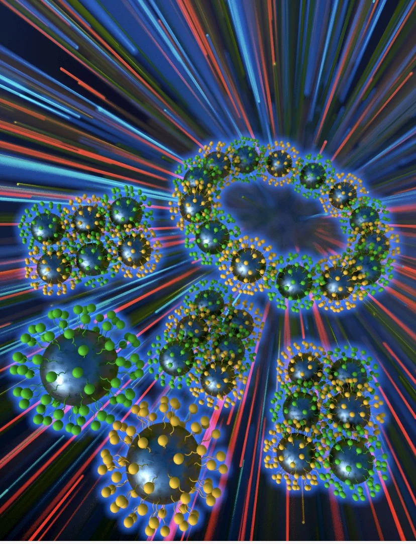
(i) Phenolic Poly(acid) Nanotherapeutics. In our lab, we employ nanoengineering principles to design structurally defined nanomedicines with improved tumor tropism and controllable therapeutic behavior. Inspired by the current poly-prodrug literature, we are extending this concept to polymer architectures derived from therapeutic acid-based monomers, enabling the formation of nanoparticles with high densities of active units and tunable, sustained bioactivity. These nanostructures also incorporate optical signatures for noninvasive tracking in vivo, supporting real-time assessment of delivery and therapeutic performances.
(ii) Organotropic LNPs. In parallel, we are developing disease-tropic lipid nanoparticles capable of selective accumulation in clinically relevant disease sites including metastatic tumors and atherosclerosis plaques. Together, these efforts contribute to a modular, programmable nanomedicine pipeline that enhances precision in both cancer therapy and image-guided interventions.
Selected Publications: (i) Rezaei, B., Harun, A. et al. Adv. Health. Mater 2024; (ii) Srivastava, I. et al.ACS Applied Materials & Interfaces 2021; (iii) Srivastava, I. et al.ACS Applied Materials & Interfaces 2020; (iv) Schwartz-Duval, A. S. et al., Nat. Comm 2020; (v) Misra, S. K., Srivastava, I., et al., Journal of American Chemical Society 2017.
Students Leading this Direction: Mahenour Megahed (PhD Student), Isabella Vasquez (Barry Goldwater Scholar), Kaylee Herrera (URS Scholar).
Collaborators: (i) Dr Ulrich Bickel (TTUHSC Pharmacy); (ii) Dr. Josh Tropp (TTU Chemistry)
Current Funding Sources:
Research Direction #4. Spectroscopic and Optical-Based Platforms
For Disease Diagnostics
In recent years, optical and surface-enhanced Raman scattering (SERS)-based spectroscopy techniques have emerged as pivotal tools in disease detection. Enhancing the sensitivity and specificity of these spectral detection methods is crucial for developing accurate diagnostic applications that can be employed at point-of-care. The integration of artificial intelligence (AI) technologies presents a significant opportunity to further improve detection accuracy through advanced machine learning algorithms. By analyzing complex spectral data, AI can identify patterns and biomarkers that may be missed by traditional methods, ultimately leading to more reliable and timely diagnosis interventions. This convergence of both optical and SERS with AI underscores our commitment to advancing diagnostic capabilities and improving patient outcomes.
To achieve this goal, our lab focuses on designing novel bioinspired optical and SERS-based nanosensors, utilizing innovative design principles to facilitate a wide range of analyte detections pertinent to biomedical health and environmental monitoring. Our research encompasses diverse applications, from identifying cancer-cell secreted exosomes in patients with solid tumors to detecting microplastics and nanoplastics in aquatic samples from the Lubbock community. Our primary emphasis in exosome studies is on cancer, particularly hepatocellular and pediatric cancers, due to the availability of patient tumor samples from our clinical collaborators at TTUHSC. We collaborate with data scientists to analyze the substantial datasets generated, identifying trends that enhance our ability to use exosomes for predicting cancer, monitoring treatment responses, and assessing cancer radioresistance. This multidisciplinary approach aims to advance diagnostic capabilities and improve patient care.
Selected Publications: (i) Vasquez, I. et al.WIRE Nanomed. 2025; (ii) Lin, L.,..,Srivastava, I.,…, Ye, J. etal. ACS Applied Materials & Interfaces 2025; (iii) Srivastava, I. et al.ACS Applied Materials & Interfaces2024; (iv) Srivastava, I. et al.ACS Nano 2022; (v) Alafeef, M., Srivastava, I. et al.ACS Sensors 2020; (vi) Srivastava, I. et al., Small Methods 2020; (vii) Pandit, S. etal., ACS Sensors 2019.
Students Leading this Direction: Md. Hasnat Rashid (PhD Student), Alan Ramirez (CISER Scholar).
Collaborators: (i) Dr. Wen Jiang (MD Anderson, Oncology); (ii) Dr. Balakrishna Koneru (TTUHSC Pediatrics); (iii) Dr. Zeeshan Ahmad (TTU ME), (iv) Dr. Danielle Levitt (TTU Kinesiology).
Current Funding Sources:
Surgical resection remains the cornerstone of solid tumor treatment. However, the ability to distinguish between malignant and healthy tissue often relies on subjective tactile and visual cues, as well as the surgeon’s individual experience. This reliance frequently results in positive surgical margins (PSM), where residual tumor tissue is inadvertently left behind. PSMs significantly degrade patient survival rates, increase the likelihood of recurrence, and pose substantial economic burdens on the healthcare system. To address these challenges, our lab is developing next-generation optical contrast agents designed to provide high-fidelity, real-time intraoperative guidance:
(i) NIR-II Fluorescence Imaging. We engineer organic polymer-based and cyanine-based nanofluorophores responsive to the Near-Infrared II (NIR-II) window(1,000–1,700 nm). Unlike the NIR-I imaging (700-900 nm) currently used in clinical intraoperative fluorescence-guided surgery (FIGS), NIR-II offers significantly reduced tissue scattering, deeper anatomical penetration, and a vastly improved signal-to-background ratio, enabling the detection of sub-millimeter buried tumor lesions, pulmonary emboli, or atherosclerosis plaques.
(ii) Afterglow Luminescence Imaging. In parallel, we leverage afterglow imaging, the detection of light emission that persists long after the cessation of external excitation. By eliminating the need for real-time light stimulation during detection, afterglow imaging effectively circumvents tissue autofluorescence and enables deep tissue imaging with ultra-high signal-to-background ratios. We develop novel organic polymers-based and cyanine-based nanofluorophores serving as highly sensitive optical beacons, allowing for the precise mapping of tumor boundaries in deep-seated tissues without the interference of background noise.
By integrating these advanced optical properties, we aim to provide surgeons with a “molecular map” that ensures complete tumor removal, minimizes adverse outcomes, and sets a new standard for precision oncology.
Selected Publications: (i) Vasquez, I. et al.ACS Nanoscience Au 2025; (ii) Bendele, N. et al.Adv. Funct. Mater 2024;
(iii) Gill, N. et al.Adv. Healthcare Mater 2024; (iv) Gill, N. et al.Adv. Optical Mater 2025.
Students Leading this Direction: Isabella Vasquez (Barry Goldwater Scholar), Asma Harun (PhD Candidate).
Collaborators: (i) Dr. Shreya Raghavan (TAMU BME); (ii) Dr. Josh Tropp (TTU Chemistry); (iii) Dr Ulrich Bickel (TTUHSC Pharmacy); (iv) Dr. Syed Muhammad Usama (UT San Antonio Chemistry); (v) Dr. Ognjen Milanjanic (University of Houston Chemistry).
Current Funding Sources:

Our lab engineers advanced tools to overcome the limitations of traditional surgical and interventional procedures. Currently, the success of both solid tumor resection and thrombolytic therapy often depends on subjective visual cues, leading to incomplete tumor removal or ineffective clot dissolution. To address these challenges, our research is organized into two primary thrusts:
(i) Precision Oncology & Surgical Imaging.To accelerate the translation of the nanofluorophores developed in our lab in Research Direction #1, we develop 3D-printed tumor-mimicking phantoms that precisely replicate the optical properties, enzyme composition, and contrast agent concentrations found in vivo, allowing for the rigorous refinement of surgical imaging tools.
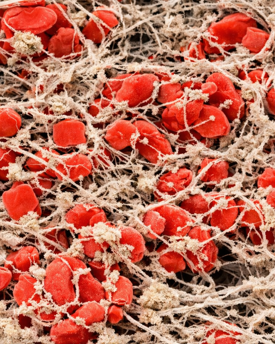
(ii) Biomimetic 3D Blood Clots for Vascular Intervention.Parallel to our oncology work, we engineer hydrogel-based 3D blood clots that serve as sophisticated in vitro benchmarks. These models are used to evaluate the efficacy of next-generation investigational thrombolytics and photothermal ablation strategies, providing a controlled environment to optimize clot dissolution and thermal distribution before clinical use.
Selected Publications: (i) Vasquez, I. et al. ACS Nanoscience Au 2025; (ii) Harun, A. et al.ACS Nano 2025;
(iii) Bendele, N. et al.Adv. Funct. Mater 2024; (iv) Srivastava, I. et al.ACS Applied Materials & Interfaces 2024;
(v) Srivastava, I. et al.ACS Nano 2023.
Students Leading this Direction: Asma Harun (PhD Candidate), Kaylee Herrera (AHA Undergrad. Asst.) Itzel De Leon (URS Scholar), Victoria Vargas (URS Scholar).
Collaborators: (i) Dr. Paul Egan (TTU ME); (ii) Dr. Anirban Sen Gupta (CWRU BME).
Current Funding Sources: